Ultra-Pro II™ In-Plane Ultrasound Needle Guides-Single-Angle
Ultra-Pro II™ In-Plane Ultrasound Needle Guides-Single-Angle
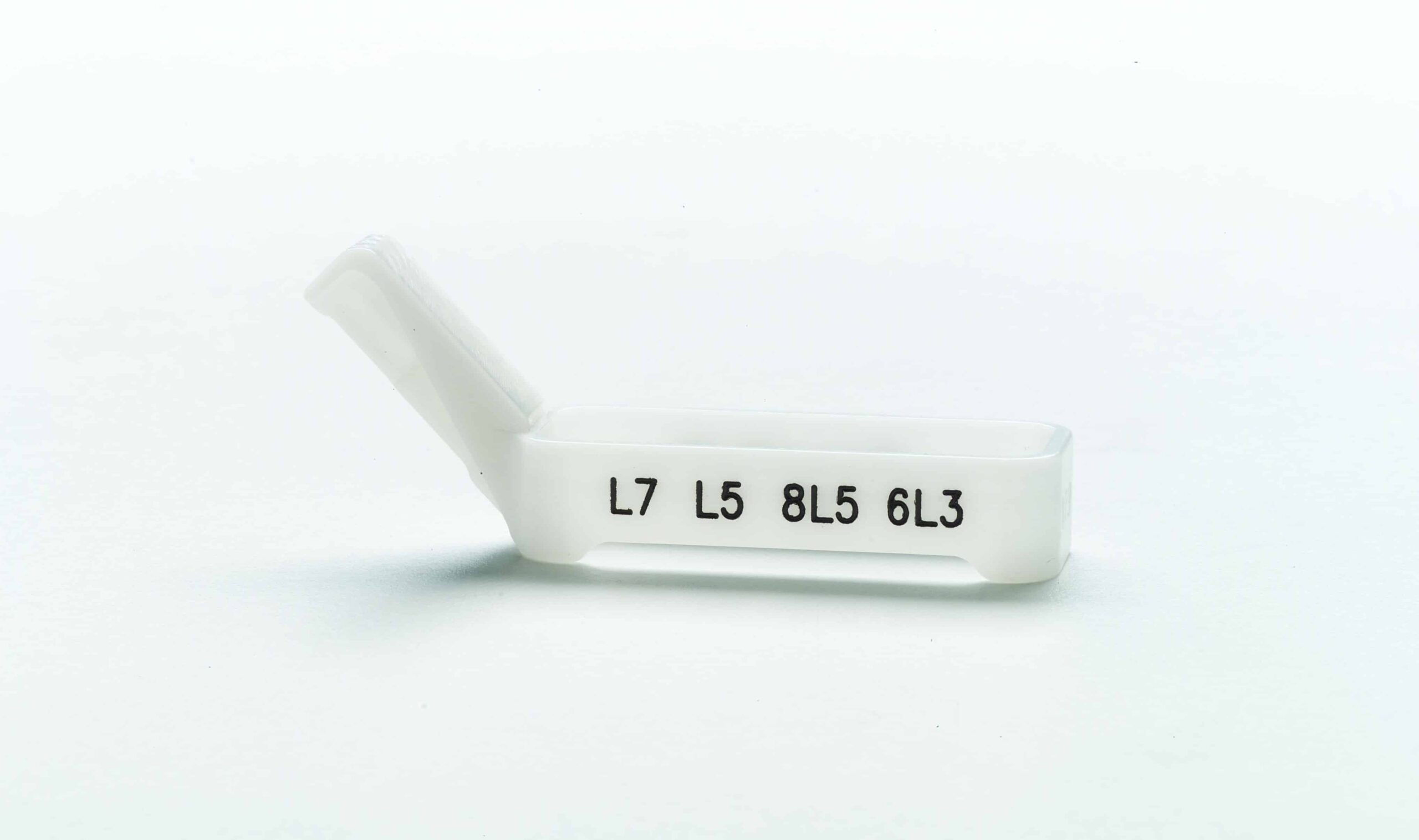
Needle Guidance System utilizes a two-part system consisting of custom reusable bracket and the disposable snap-on needle guide called Ultra-Pro II. Single-angle brackets offer imaging professionals a fixed angle needle path during puncture procedures.
Needle Guidance System utilizes a two-part system consisting of custom reusable bracket and the disposable snap-on needle guide called Ultra-Pro II. Single-angle brackets offer imaging professionals a fixed angle needle path during puncture procedures. The Ultra-Pro II needle guide incorporates flexibility and advanced features to increase productivity during ultrasound-guided procedures. The Ultra-Pro II guide features a large tab for improving quick-release function, allowing easy detachment of the needle from the transducer. Easy-to-read gauge sizes on the inserts make it simple to identify and alter gauge size in a darkened ultrasound suite. The guide features a large funnel for instrument insertion and will accept a range of sizes including: 8.5FR, 14-23GA (19 GA not available). Brackets should be cleaned and disinfected according to your User's Guide.
Downloads
Clinical Benefits
Ultra-Pro II™ Needle Guide
Accurate instrument placement with reduced procedure time resulting in adequate yield for pathology.
- Increased clinical confidence with improved needle visualization during real-time imaging1,2
- Improves needle visualization within the tissue which increases patient safety5
- Reduced procedure time from a guided biopsy will correlate with reduced needle manipulation and hence less tissue trauma and less patient discomfort.2
- Improved time efficiency by increasing needle visualization and reducing number of needle passes.2,3, 5,6
- Efficient and reproducible biopsy technique compared to freehand, ensures confident outcomes.2
- Decreases the number of passes required for adequate tissue yield and lowers the risk of developing major complications by 305% compared to freehand during renal biopsy.3
- Improve patient safety and comfort during peripheral nerve blocks.4
Sources
- Phal P, Brooks M, Wolfe R. “Sonographically Guided Biopsy of Focal Lesions: A Comparison of Freehand and Probe Guided Techniques using a Phantom.” American Journal of Roentgenology (AJR). 2005; 184:1652-1656.
- Shabana, W, Kielar A, Vermani V, Fernandes D, Antoniscu R, Schweitzer M. “Accuracy of Sonographically Guided Biopsy Using a Freehand Versus Needle Guided Technique Computer Tomographic Correlation Study.” Journal of Ultrasound in Medicine (JUM). 2013; 32: 535-540.
- Prasad N, Kumar S, Manjunath R, Bhadauria D, Kaul A, Sharma R, Gupta A, Lai H. Jain M, Agrawal V. “Real-time Ultrasound-Guided Percutaneous Renal Biopsy with Needle Guide by Nephrologists Decrease Post-Biopsy Complications.” Clinical Kidney Journal. 2015: 8: 151-156.
- Bluvol N, Korneck A, Shaikh A, Del Rey Fernandez D, Taves D, Fenster A. “Freehand Versus Guided Breast Biopsy: Comparison of Accuracy, Needle Motion and Biopsy Time in a Tissue 6odel.” American Journal of Roentgenology (AJR). 2009; 192:1720-1725.
- Clendenen S, Robards C, Greengrass R. “Popliteal Catheter Placement Utilizing Ultrasound Needle Guidance System.” Local and Regional Anesthesia. July 28, 2010; 3:45-48.
- Mansour MA, Sonbaty M. “Enhancing Needle Visualization During Parasagittal Approach in Paravertebral Blocks for Patients Undergoing Simple Mastectomy Using In-Plane, Multiangle Ultrasound Needle Guidance System.” Saudi Journal of Anesthesia. 2016; 10:33-37.
Related products
-
Ultra-Pro e™ In-Plane Ultrasound Needle Guides
Ask for quote -

Orchid Spec Medium
Ask for quote This product has multiple variants. The options may be chosen on the product page -

Virgo SPX insert
Ask for quote